
FIG. 4.
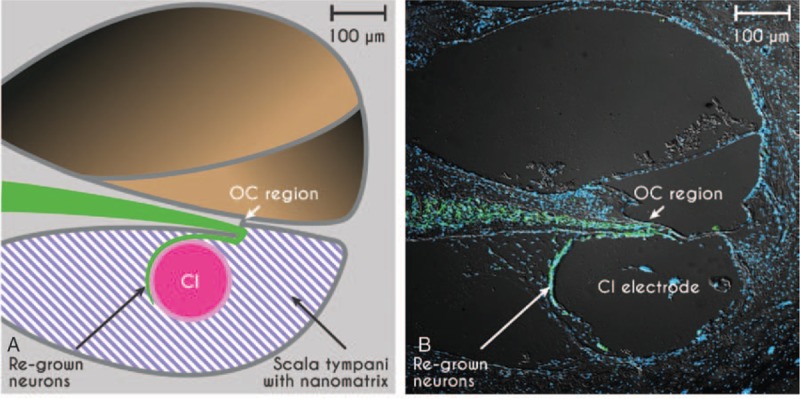
Proof of concept of the gapless interface between auditory neurons (green) and the CI-electrode array in scheme (A) and in the deafened guinea pig inner ear in vivo (B). Auditory neurons are stained with a neuron-specific marker for β-III tubulin (green, TUJ), cell nuclei are stained in blue (DAPI nuclear staining), photograph provided by Marcus Müller and Hubert Löwenheim. CI indicates cochlear implants.