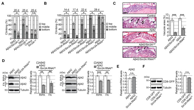
Figure 2. Neuronal knockdown of Sin3A protects against chronic ER proteinopathy in Aβ42 fly brains.

(A–B) Heterozygous loss-of-function mutation (Sin3ALOF) or RNAi-mediated knockdown (Sin3A RNAiGD and Sin3A RNAiKK) of Sin3A suppressed Aβ42-induced locomotor defects. Mean ± SD, n = 5, *p < 0.05 by Mann–Whitney U test. n.s.: not significant. (C) Sin3ALOF or Sin3A RNAi suppressed Aβ42-induced neurodegeneration. Percentages of vacuole areas in fly brain cortices are shown (indicated by arrows). Scale bar: 10 μm. Mean ± SEM, n = 7–12 hemispheres, ***p < 0.001 by Student's t-test. (D) RNAi-mediated knockdown of Sin3A in neurons reduced Aβ42 levels in fly brains. Detergent-soluble (RIPA) and -insoluble (FA) fractions from heads were analyzed by western blotting with anti-Aβ antibody. Mean ± SEM, n = 3–4. **p < 0.01 and ***p < 0.001 by Student's t-test. (E) Neuronal knockdown of Sin3A did not alter Aβ42 mRNA levels in fly brains, as determined by qRT-PCR. Mean ± SEM, n = 3–4, n.s.: not significant (Student's t-test). (F) Neuronal knockdown of Sin3A did not reduce levels of CD8-GFP protein. Mean ± SEM, n = 3, n.s.: not significant (Student's t-test). See also Figure S2 and Table S5.
