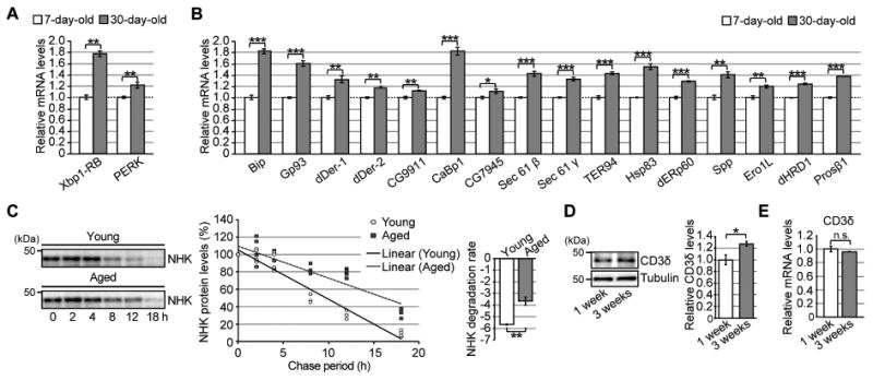
Figure 6. Expression levels of UPR-related genes are elevated, whereas ERAD activity is reduced, in aged fly brains.

(A) mRNA expression levels of Xbp-1RB and PERK were elevated in aged fly brains. (B) Age-dependent increases in mRNA levels of UPR-related genes downstream of Ire1/Xbp1 in fly brains. For (A–B), mRNA levels in the heads of 7- or 30-day-old flies were analyzed by qRT-PCR. Open bars: 7 days old; filled bars: 30 days old. Mean ± SEM, n = 4–8, *p < 0.05, **p < 0.01, and ***p < 0.001 by Student's t-test. (C) (Left) Degradation of the ERAD substrate NHK was significantly slower in aged fly brains. After induction of NHK expression for 2 days, the decay rate of NHK protein in fly brains was analyzed for up to 18 hours by western blotting. (Right) Degradation rates. Mean ± SEM, n = 4, **p < 0.01 by Student's t-test. (D) Age-dependent increases in an ERAD substrate protein, CD3δ-YFP, in fly brains. Heads of flies expressing CD3δ-YFP at the age of 1 or 3 weeks were analyzed by western blotting. Mean ± SEM, n = 3, *p < 0.05 by Student's t-test. (E) mRNA levels of the CD3δ transgene in fly brains were not altered by age, as determined by qRT-PCR. Mean ± SEM, n = 4, n.s.: not significant (Student's t-test). See also Tables S3–5.
