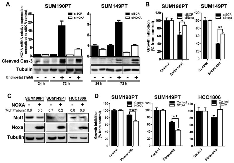
Figure 4. NOXA expression plays an important role in the regulation of sensitization of TNBC and IBC cells to treatment.
SUM190PT and SUM149PT cells were transfected with NOXA (siNOXA) or Scrambled (siSCR) siRNA through electroporation. Knockdown of NOXA mRNA and induction of apoptosis as measured by cleaved caspase-3 after siRNA inhibition were confirmed by quantitative RT-PCR and immunoblotting analysis (A), respectively, after entinostat treatment for 24 and 72 hours. Cell proliferation after siRNA and entinostat treatment was measured by SRB staining after 72 hours (B). SUM190PT, SUM149PT, and HCC1806 cells were transfected with either a NOXA-expressing vector or empty control vector by electroporation. Expression of NOXA, as well as MCL1, was analyzed by immunoblotting analysis 72 hours after transfection (C). Pixel density of MCL1 was quantified for each condition, and the ratios of MCL1/tubulin are shown above the blots; tubulin expression was used as a protein loading control. Proliferation of cells with NOXA overexpression in response to treatment with pimasertib (2.5 μM) was determined by SRB staining after 72 hours (D). Data were pooled from three independent experiments and presented as mean ± SEM. *, P < 0.05; **, P < 0.005; ***, P < 0.0001.

