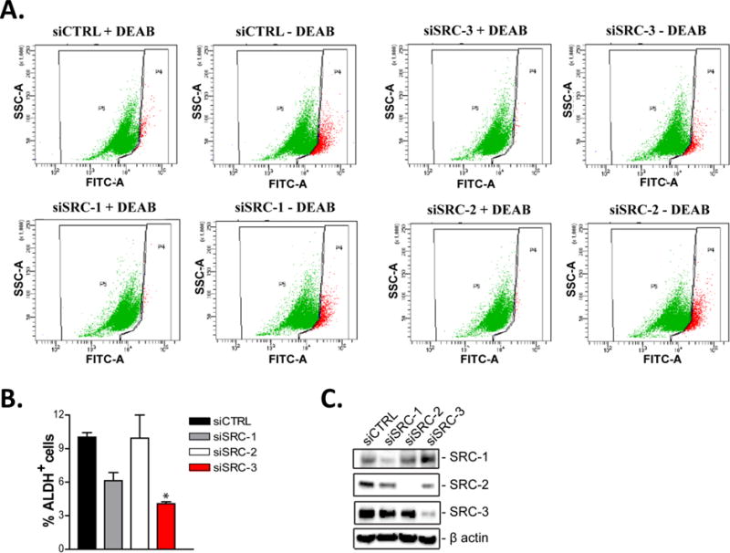
Figure 2. SRC-3 regulates the proportion of CSCs.

Decreased expression of SRC-3 reduces the % ALDH+ cell population. SKBR3 cells were transiently transfected with either control (siCTRL), SRC-1 (siSRC-1), SRC-2 (siSRC-2) or SRC-3 (siSRC-3) targeting siRNAs for 48 hours. (A-B) Cells were then assayed for ALDH activity using flow cytometry. Representative flow cytometry dot plots are shown. The graphed data represents the mean ± S.E.M. (n=3). One-way ANOVA and Tukey’s multiple comparison tests were used for significance testing. * = p < 0.05. (C) 48 hours post transfection total protein was isolated from the cells and subjected to immunoblot analysis with antibodies to SRC-1, SRC-2, SRC-3 and β-actin.
