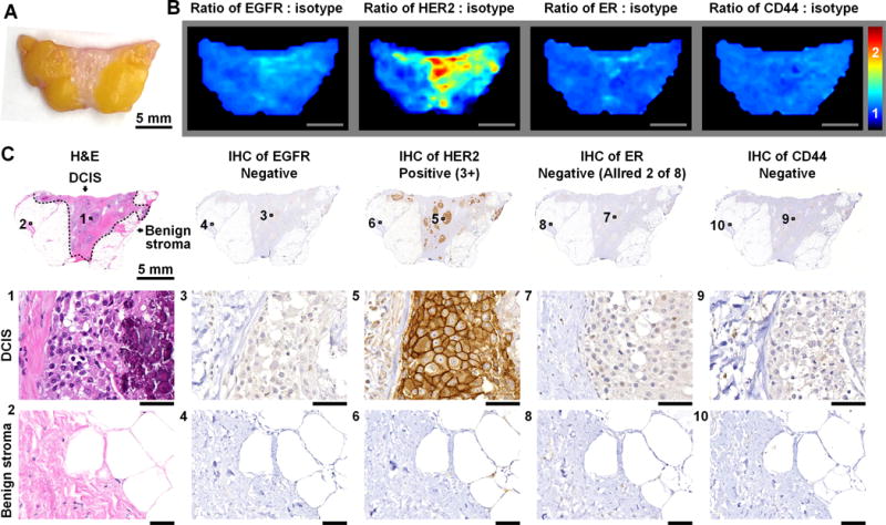
Figure 2. REMI enables multiplexed detection of disease-associated biomarkers.

Here, an example is shown of a patient with a HER2-positive neoplasm in which REMI successfully identifies the over expression of this cell-surface biomarker. A, Photograph of a human breast specimen with DCIS. B, REMI results. Unlabeled scale bars represent 5 mm. The color bar indicates NP ratios. C, Validation data: H&E histology and immunohistochemistry (IHC). H&E histology is the clinical gold-standard method for the detection of carcinoma, and IHC is a clinical gold-standard method for the assessment of protein expression. In this example, the specimen is positive for HER2 and negative for ER, EGFR and CD44. Unlabeled scale bars represent 50 μm.
