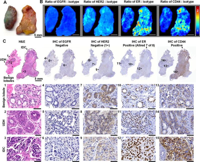
Figure 3. REMI enables discrimination between benign and malignant lesions.

A, Photograph of two tissue specimens from a single patient. The specimen on the left presents regions of usual ductal hyperplasia (UDH, benign lesion), and the specimen on the right presents invasive ductal carcinoma (IDC, malignant lesion). B, REMI reveals that the overexpression of ER and CD44 is associated with IDC (specimen on right) but not UDH (specimen on left). Unlabeled scale bars represent 5 mm. The color bar indicates NP ratios. C, Validation data: H&E and IHC. The specimen with IDC is positive for ER and CD44, which is concordant with the REMI results. The specimen with UDH is negative for all four biomarkers. See text for details on how the IHC results are scored based on standard-of-care methods. Unlabeled scale bars represent 50 μm.
