
Figure 4. The multiplexed imaging of multiple biomarkers, enabled by REMI, improves detection sensitivity for malignancies with molecular phenotypes that are spatially and/or temporally varying.
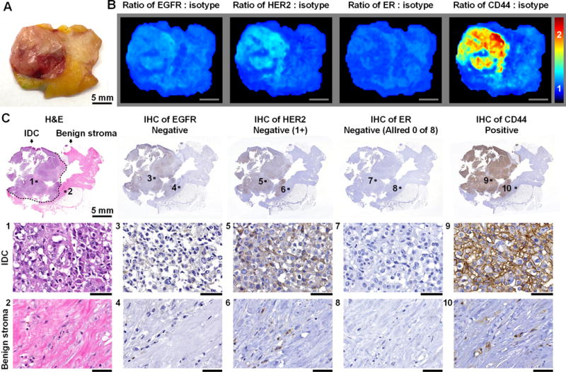
A, Photograph of a human breast specimen with IDC that was ER-positive on biopsy but ER-negative on lumpectomy, presumably as a result of neoadjuvant endocrine therapy. B, REMI results. Unlabeled scale bars represent 5 mm. The color bar indicates NP ratios. C, Validation data: H&E and IHC. The specimen is positive for CD44 and negative for HER2 (cytoplasmic staining only), ER and EGFR. See text for details on how the IHC results are scored based on standard-of-care methods. Unlabeled scale bars represent 50 μm.