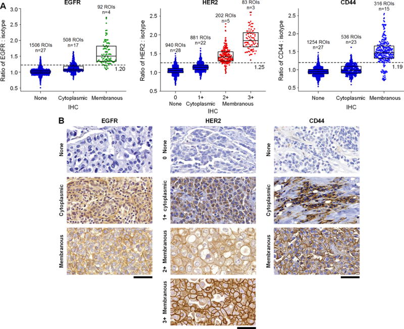
Figure 6. REMI can quantify the expression of cell-surface biomarkers but is intrinsically insensitive to intracellular (e.g. cytoplasmic and nuclear) expression.

A challenging issue for pathologists, when assessing the expression of cell-surface biomarkers with IHC, is differentiating between intracellular and membranous staining and only using the latter as a basis for scoring the expression levels of cell-surface proteins. REMI stains and images fresh tissue surfaces that are composed of mostly intact cells, in which the large SERS NPs are not easily internalized. Therefore, REMI is intrinsically less sensitive to intracellular targets and provides reliable quantification of cell-surface biomarker expression, with good correlation to IHC scores (as determined by expert pathologists). A, Correlation between REMI and IHC results for cytoplasmic vs. cell-surface membrane targets (2106 ROIs in total). B, Examples of IHC images showing nonexistent or weak cytoplasmic staining (row 1), intermediate or strong cytoplasmic staining (row 2) and cell-membrane staining (rows 3 and 4). Unlabeled scale bars represent 50 μm. The box plots in panel A are all significantly different from each other (p < 0.001).
