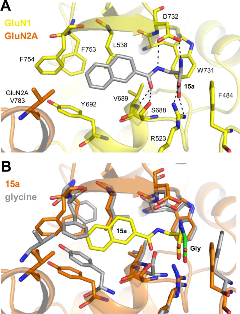
Figure 3.
Ligand-docking and molecular dynamics simulations with 15a in the GluN1/2A ABD heterodimer structure. (A) Representative stable binding mode of 15a adopted in four molecular dynamics simulations using two high scoring poses from ligand-docking into the structure of the GluN1/2A ABD dimer (see the Supporting Information for additional figures and details on the molecular modeling). (B) Overlay of the structure from molecular dynamics simulations with bound 15a (protein in orange; 15a in yellow) and the GluN1/2A ABD crystal structure with bound Gly (protein in gray; Gly in green).

