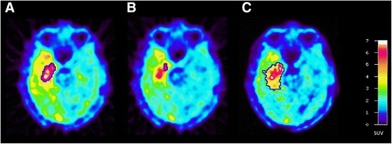
Fig. 3.

ROI definition in a patient with a glioblastoma in the right temporal lobe. a The tumour volume as delineated by TBR > 1.8 based on data reconstruction of centre A. b The 90% isocontour of the 10–30-min image for TAC generation in centre A and c the tumour volume as delineated by a TBR > 1.6 in centre B which is also used TAC generation
