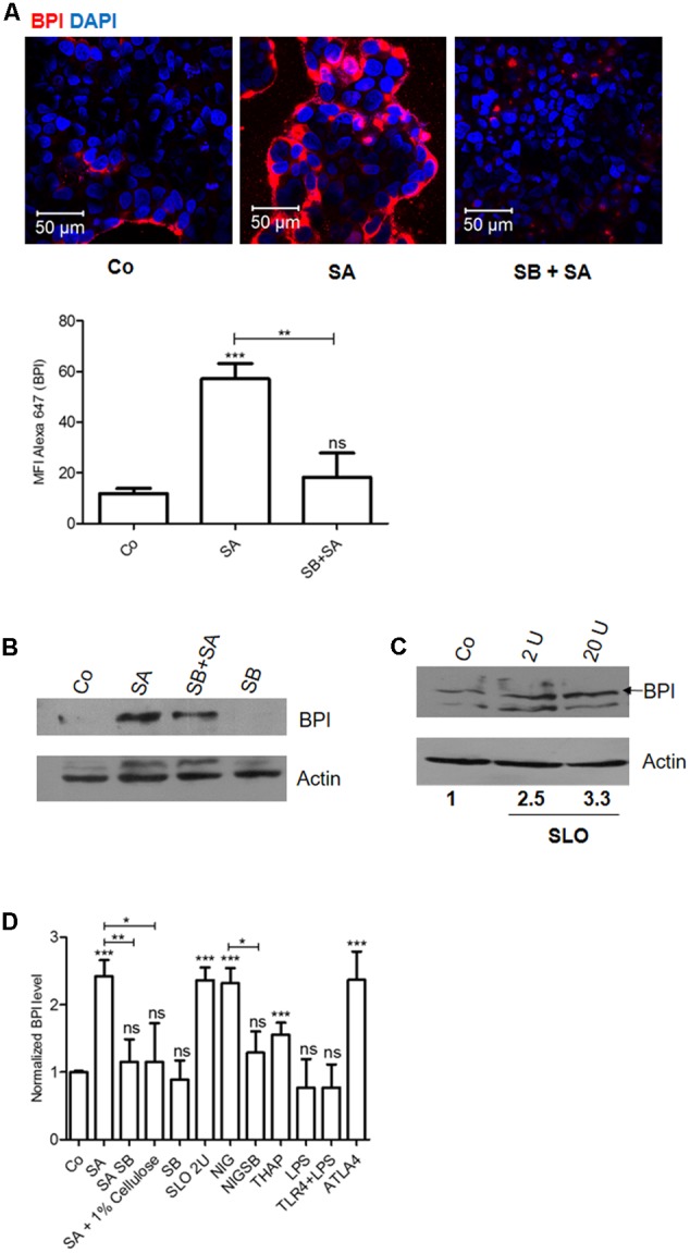
FIGURE 2.

Caco-2 cells recognize Staphylococcus aureus induced stress and increase BPI expression in a p38 dependent manner. Caco-2 cell monolayers were either left untreated or treated with SB203580 (p38-MAPK inhibitor, SB) 1 h before S. aureus (SA) infection. (A) Cells were fixed 24 h post-infection and were immunostained with anti-BPI antibody. Representative images are shown. Bottom: The Mean Fluorescent Intensity (MFI) of BPI was calculated using Zen software and plotted. (B) Cells were lysed 24 h post-infection and BPI levels were checked by western blot. (n = 4 experiments) Co (media alone). (C) Cells were treated with Streptolysin O (2, 20 units) for 30 min and BPI levels were checked by western blot 24 h post-treatment. (n = 3 experiments). (D) BPI protein levels in Caco-2 cells were normalized to β actin internal control and expressed relative to medium alone control. The data is expressed as relative BPI levels (x-axis; n = 4 experiments). Statistical analysis was done by the students’ t-test. Key: ∗∗∗p < 0.001, ∗∗p < 0.005, ∗p < 0.05, ns = not significant.
