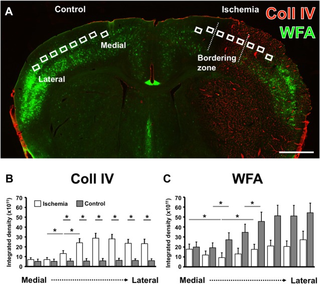
Figure 3.

Comparative semi-quantitative analyses addressing immunoreactivity for Coll IV- and WFA-staining in the neocortex of mice. A coronal brain section illustrates the selected regions of interest (ROIs) used for semi-quantitative analysis in the ischemia-affected hemisphere and in the corresponding contralateral area that served as control (A). The semi-quantitative comparison of the Coll IV-immunoreactivity (B) and WFA-staining (C) between both hemispheres (gray bars indicate the contralateral and white bars the ischemia-affected hemisphere) shows a significant up-regulation of the Coll IV-fluorescence signal that corresponds with a relatively reduced lectin-histochemical signal for WFA alongside from the medial to the lateral part of the neocortex. Bars indicate mean values, and added lines stand for the standard error of mean. Horizontal lines with added significance levels represent inter-hemispheric comparisons (short lines) as well as intra-hemispheric comparisons (long lines). *p < 0.05. Scale bar = 1 mm.
