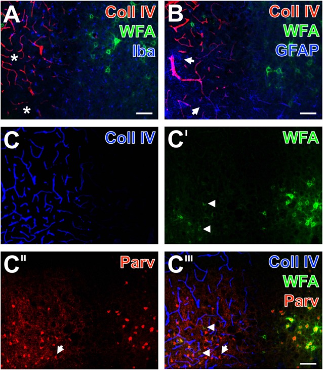
Figure 4.

Infarcted mouse: up-regulated Coll IV and decomposed PNs in ischemic neocortex—allocated with microglia/macrophages, activated astroglia and altered parvalbumin-immunolabeling. The ischemic border zone in the somatosensory cortex (A,B) exhibits nearly complementary distribution patterns for WFA (green) and strongly up-regulated Coll IV (red) 1 day after induction of ischemia. Additionally, (A) displays in the apparently non-affected zone evenly distributed ionized calcium binding adaptor molecule 1 (Iba)-positive microglia (Cy5, color-coded in blue), whereas the infarcted zone is partly devoid of Iba-immunoreactivity (asterisks). In parallel, glial fibrillary acidic protein (GFAP)-immunolabeling (blue) in (B) reveals also fragmented astrocytes (arrows) in the damaged tissue. (C–C‴) show concomitant staining of Coll IV, WFA and parvalbumin as markers for most neocortical net-bearing neurons. Up-regulated Coll IV-immunoreactivity (C) indicates the ischemia-affected tissue, where WFA-stained nets appear decomposed (arrowheads in C′ and the merged picture C‴) or even abolished. Parvalbumin-immunolabeling (C″,C‴) visualizes shrunken neurons (exemplified by an arrow) and enhanced neuropil staining in the same area. Here, the overlay (C‴) elucidates only a few PNs around parvalbumin-containing neurons. Scale bar in A,B,C‴ (also valid for C–C″) = 75 μm.
