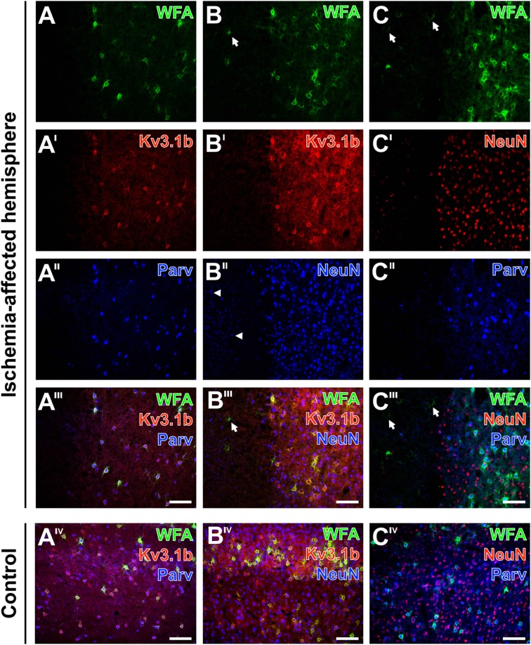
Figure 9.
Alterations of parvalbumin, Kv3.1b and neuronal nuclei (NeuN) in neurons surrounded by ischemia-affected, WFA-stainable nets in mouse neocortex. One day after stroke induction PNs at the ischemic border zone in the neocortex are revealed by WFA-staining (green) in combination with the immunolabeling of (A–A‴) Kv3.1b + parvalbumin, (B–B‴) Kv3.1b + NeuN and (C–C‴) NeuN + parvalbumin. Evenly distributed PNs in the apparently healthy tissue contrast to the ischemia-affected area (A) devoid of WFA-labeled structures or (B,C) with only some remnants of nets (arrows in B,B‴,C,C‴). Kv3.1b and parvalbumin are co-localized in all detected neurons (A′–A‴) which results in the pink appearance of neurons co-expressing both markers (A‴). While Kv3.1b and NeuN-immunolabeling appear both unaltered in the sharply delineated healthy tissue, the ischemia-affected region additionally contains structures with diminished NeuN-immunosignals (arrowheads in B″). Additionally, NeuN-staining (C′) is also less affected in comparison to parvalbumin-immunoreactivity which remains restricted to coarse neuropil staining in the injured tissue (C″). Merged images show all three marker combinations both in the ischemia-affected neocortex (A‴–C‴) and in the same neuroanatomical area on the contralateral, non-affected side (AIV–CIV). Scale bars A‴,AIV,B‴,BIV,C‴,CIV (also valid for all other micrographs) = 100 μm.

