
Figure 3.
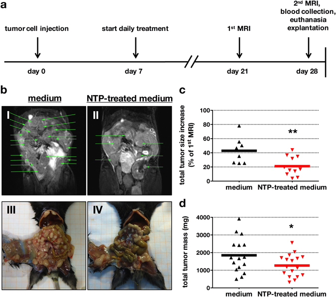
NTP-treated medium decreased number, growth, and size of pancreatic lesions in vivo. The experimental timeline (a) is given. The number of tumor nodes in the peritoneum was followed by MRI and was elevated in the control animals (I) compared to NTP-treatment group (II) at the end of treatment (d28). Also, corresponding macroscopic findings for control (III) and NTP-treated (IV) animals are shown (b). MRI-based calculation of tumor volume revealed a significantly decreased total tumor growth in the treatment group (c). At day 28, animals were sacrificed and tumor nodes were excised and weighed, showing a significantly decreased total tumor mass (d). Representative images of 13 animals are given, green arrows indicate tumor nodes (one green arrow per lesion) (b). Data are presented as mean of 8–17 animals (c,d).