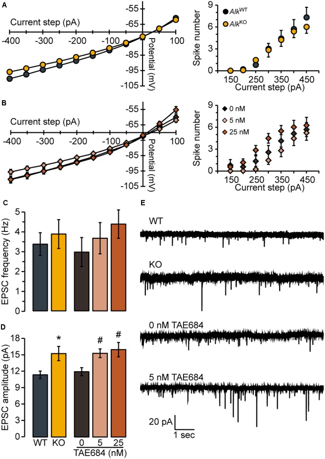
FIGURE 2.

Loss of ALK does not alter membrane properties or excitability but promotes excitatory synaptic transmission. Membrane voltage (left) and spike firing (right) responses to hyperpolarizing and depolarizing current steps (300 ms duration) for AlkKO vs. AlkWT (A) or TAE684 (5 and 25 nM) vs. vehicle (0 nM) (B). Frequency (C) and amplitude (D) of spontaneous EPSCs in AlkWT and AlkKO, and in vehicle-treated (0 nM) and TAE684-treated (5 and 25 nM). ∗p = 0.02, AlkKO vs. AlkWT. #p = 0.02, 5 nM vs. 0 nM, and p = 0.04, 25 nM vs. 0 nM (Games-Howell post hoc comparisons). (A–D) Data are represented as group means ( ± SEM) for AlkWT (n = 10 cells, four mice), AlkKO (n = 13 cells, five mice), vehicle-treated (n = 8 cells, five mice), 5 nM TAE684 (n = 9 cells, five mice), and 25 nM TAE684 (n = 14 cells, six mice). (E) Representative current traces showing NAcSh D1MSN spontaneous EPSCs.
