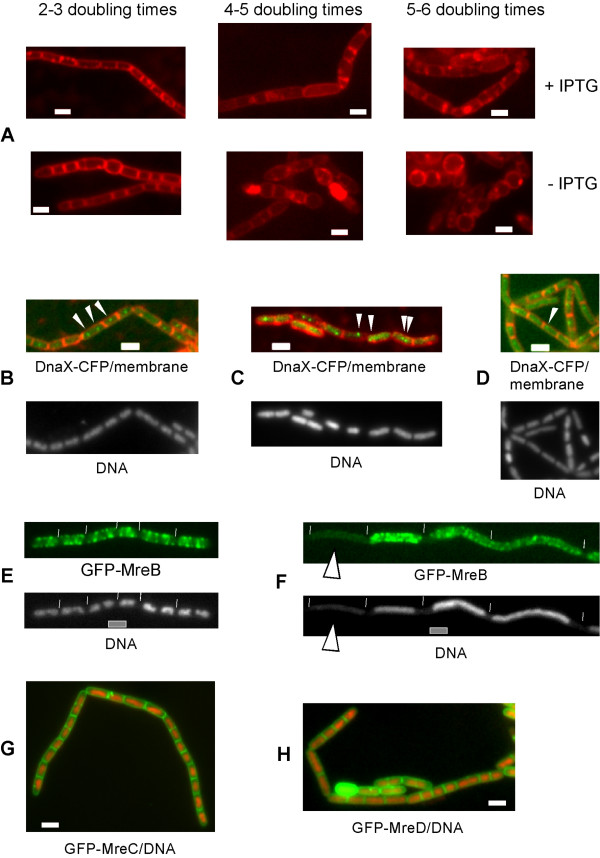
Figure 2.
Depletion of MreB in the presence of MreC and of MreD affects both cell shape and segregation of chromosomes, and affects localization of the replication factory. Fluorescence microscopy of exponentially growing Bacillus subtilis cells. A) MreB is depleted in the presence (+IPTG, upper panels) or in the absence (-IPTG, lower panels) of MreC and MreD, 2–3, 4–5 and 5–6 doubling times indicate the time after the onset of depletion. B) Localization of DnaX-CFP in wild type cells, or C) 2–3 doubling times after depletion of MreB, or D) 2–3 doubling times after depletion of Mbl. Arrowheads indicate the proper positioning of DnaX-CFP in wild type cells, and its abnormal loacalization during depletion of actin orthologs. E) Localization of GFP-MreB in wild type cells, or F) in smc mutant cells, G) localization of GFP-MreC, overlay of GFP-MreC (green) and DNA stain (red), H) localization of MreD, overlay of GFP-MreD (green) and DNA (red). White lines indicate ends of cells, white bars 2 μm.

