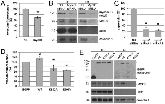
Figure 2.
Effects of myosin IC and isoform A on exosome and metalloprotease secretion by prostate cancer cells. (A) Fluorescence of the secreted exosomal fraction (stained with membrane dye Vybrant DiI) from PC3 cells transfected with nonspecific (NS) and myosin IC-targeted (myoIC) siRNA. Data were normalized to the control (nonspecific siRNA treatment) in each experiment. N = 8 cell cultures per group. Experiment was repeated 4 times. Error bar: 95% c.i. on the mean. (B). Western blot analysis of total cell (TC) extract and secreted exosomes (Ex) from the siRNA knockdown experiments. (C) Quantification of the knockdown. N = 2 cell cultures per group. Experiment was repeated 2 times. Error bar: 95% c.i. on the mean. Two different sequences were targeted by siRNA 1 and 2; siRNA 1 was used in all other experiments shown. *Significant (p < 0.0001) deviations from the control. (D) Fluorescence of the secreted exosomal fraction measured as in A, from PC3 cells transfected with EGFP fusion constructs of myosin IC isoform A. EGFP, control transfection with EGFP alone. WT, wild-type isoform A. K892A, lipid-domain substitution. E391V, motor-domain substitution. Data presentation as in (A). N = 5 cell cultures per group. Experiment repeated 5 times. Each p-value in pairwise comparisons with the control is <0.0001 (*), and all null hypotheses are rejected at familywise significance level 0.05 if regarded as a multiple comparison using the Holm-Bonferroni correction. (E) Western blot analysis of total cell (TC) extract and secreted exosomes (Ex) from the EGFP fusion experiments.

