Figure 3.

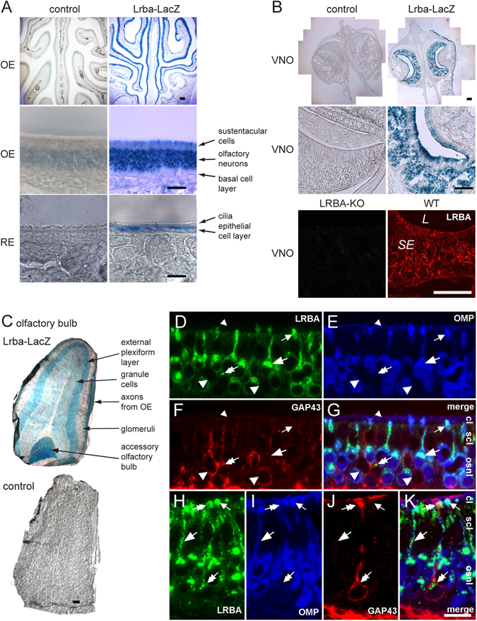
Histological and subcellular distribution of LRBA in olfactory and vomeronasal tissues. (A) LacZ enzyme histochemistry of Lrba/gt gene-trap mice (age, 3 weeks) demonstrates expression of the LRBA gene throughout the OE (top). Larger magnification (middle) shows that the staining is predominantly localized to the OSNs. Respiratory epithelium (RE, bottom) is also stained, though weaker than OE. (B) β-galactosidase staining was also seen in VNO neuron somata and the microvillar layer of Lrba/gt mice (top, middle), and confirmed by LRBA-IF of adult WT tissue (bottom; SE, sensory epithelium; L, lumen). Scale bars in A,B: overviews 100 µm, close-ups 25 µm. (C) LacZ staining of the olfactory bulb required longer incubation (16 h) than of the OE and VNO (2 h). Scale bar in C: 100 µm. (D–K) Triple immunostaining for LRBA (D,H), OMP (E,I), GAP43 (F,J) and merged images (G,K) in different regions of the adult mouse OE of low (D–G) or high (H–K) thickness. LRBA-immunoreactivity (ir) is found in the apical somatic cytoplasm and in dendrites as well as dendritic knobs of virtually all OMP-ir mature OSNs. Large and small arrows point to select LRBA-/OMP-ir cell bodies/dendrites and dendritic knobs, respectively, but LRBA is lacking in the OMP-ir ciliary layer (small arrowheads in D–G). GAP43-ir immature neurons contain no (large arrowheads in D–G) or only small amounts (double arrows in D–K) of LRBA-immunoreactivity in the apical cytoplasm of their cell bodies and in dendritic knobs. Scale bar in K for D–K: 10 µm. OE strata are designated in panels G and K: cl, cilial layer; scl, sustentacular cell layer; osnl, OSN layer.
