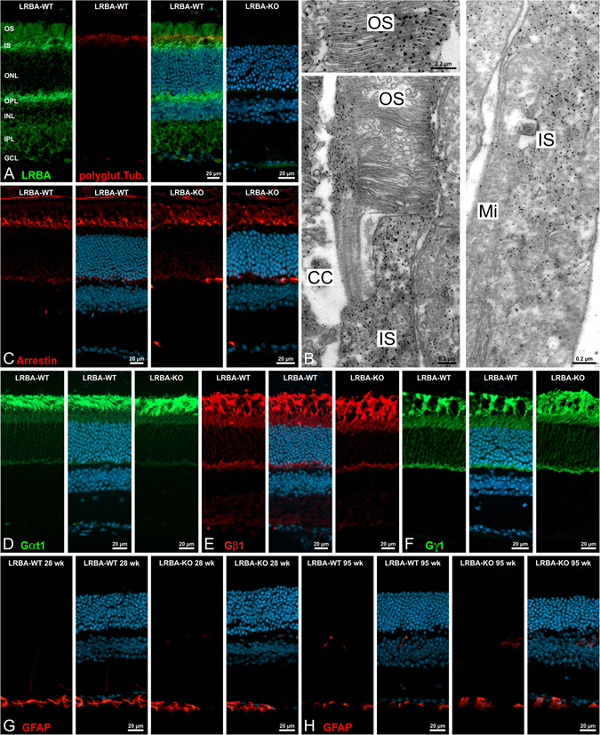
Figure 6.

Localization of LRBA and histological analysis of the LRBA-KO in the mouse retina. (A) Micrographs of a vertical cryostat section through an adult WT mouse retina triple-labeled for LRBA (green), glutamylated tubulin (marker for connecting cilia, red) and DAPI (cell nuclei, blue). LRBA-ir is prominent in the outer segment (OS), the inner segment (IS) and the outer plexiform layers (OPL), and is abolished in KO mouse retina. ONL: outer nuclear layer; INL: inner nuclear layer; IPL: inner plexiform layer; GCL: ganglion cell layer. (B) Electron micrographs showing the ultrastructural localization of LRBA-ir (pre-embedding labeling) in the cytosol of outer and inner segments of rod photoreceptors. CC: connecting cilium; Mi: mitochondria. (C) Cryostat sections through light-adapted (2 h, 150–200 lux) WT and LRBA-KO retinae were stained with anti-arrestin (red) and DAPI (cell nuclei, blue). No differences were seen between the localizations of arrestin in the WT and LRBA-KO retinae. (D–F) Cryostat sections through light-adapted (2 h, 150–200 lux) WT and LRBA-KO retinae were stained with antibodies against the subunits αt1 (green), β1 (red) and γ1 (green) of transducin. Again, no differences were detected between the localizations in the WT and LRBA-KO retinae. (G–H) WT and LRBA-KO retinae of 28 and 95 week (wk) old mice were double-labeled for GFAP (green) and DAPI (blue). No up-regulation of GFAP in retinal Müller cells as an early indicator of retinal stress was detected.
