Figure 4.

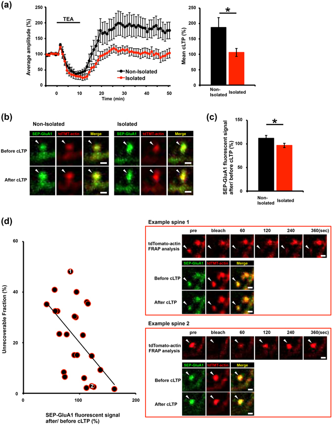
Disrupted GluA1 trafficking to the surface of spines in socially isolated rats is correlated with the increase in the stable actin fraction. (a) LTP was successfully induced in the barrel cortex of non-isolated rats (n = 13) by 10 min TEA treatment (black line). In contrast, chemical LTP (cLTP) induction was prevented in the barrel cortex of isolated rats (n = 10). (b) Representative images showing SEP-GluA1 and tdTomato-actin fluorescence on spines before and after TEA treatment in the barrel cortex of isolated or non-isolated rats. (c) Quantification of the changes in the SEP-GluA1 spine/dendrite ratio after TEA treatment. The integrated intensity of SEP-GluA1 fluorescent signal after/before cLTP on the spines was increased in non-isolated controls (n = 80) but not in isolated rats (n = 70). (d) Negative correlation between the surface expression of SEP-GluA1 at spines and the unrecoverable actin fraction at spines in the barrel cortex slices obtained from isolated rats. Two representative spine images: one with a high unrecoverable actin fraction and no cLTP-induced increase in the surface expression of SEP-GluA1 (Example spine 1) and a second with a low unrecoverable actin fraction and a cLTP-induced increase in the spine surface expression of SEP-GluA1 (Example spine 2). The scatter plot shows the changes in SEP-GluA1 expression (presented as a spine/dendrite ratio) after/before TEA treatment versus the fraction of unrecoverable tdTomato-actin at each of the individual spines. The fraction of unrecoverable actin was negatively correlated with the enhancement in surface GluA1 expression after/before cLTP in the barrel cortex slices obtained from isolated rats (r = 0.536, P < 0.05, n = 24). Representative images shown in the figures were Gaussian- and unsharp mask-filtered. Arrowheads indicate dendritic spines. Scale bar represents 1 µm. *P < 0.05 (Unpaired Student’s t-test). Error bars represent SEM.
