Figure 4.

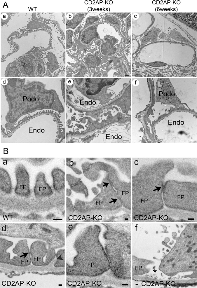
Podocyte morphology in WT and CD2AP deficient mice by TEM. (A) Representative TEM images of WT (a, d), 3 week-old Cd2ap-KO (b, e) and 6 week-old Cd2ap-KO (c, f) kidneys. WT glomeruli (a, d) show well-organized interdigitated foot processes lining up along the capillary wall with uniformly spaced slit diaphragms (SDs) between adjacent foot processes. Glomeruli of both 3 week- (b, e) and 6 week-old (c, f) Cd2ap-KO mice show massive foot process effacement, which is associated with the disappearance of the filtration slit and SDs between foot processes. Intracellular junction-like structures are frequently formed between effaced podocytes. Scale bars, 1 μm in upper panels; 500 nm in lower panels. (B) Representative TEM images of podocyte slit pores in WT (a) and Cd2ap-KO (b–f) mice. Intact slit pores are seen in WT mice (a). In Cd2ap-KO mice, narrow slit pores are frequently seen (b–e) with some cell contact area (black arrows). GBM expansion into the space between foot processes is observed (d, arrowheads). A widened slit pore with no SD is also observed (f, asterisk). Scale bar, 100 nm. Podo, podocyte; FP, foot process; Endo, endocapillary space.
