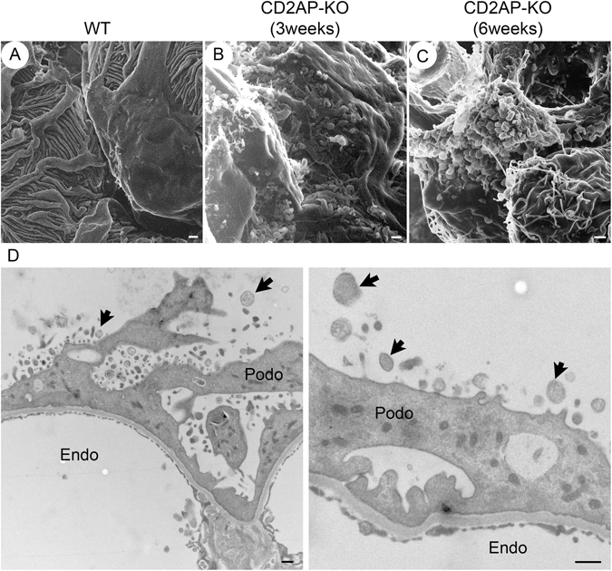
Figure 5.

Bleb-like microprojections from podocytes in CD2AP deficient mice. HIM images of podocyte cell body in WT (A), 3 week-old Cd2ap-KO (B), and 6 week-old Cd2ap-KO mice (C). The image of WT podocytes (A) shows a smooth podocyte cell body and well-organized interdigitating foot processes. Images of 3 and 6 week-old Cd2ap-KO podocytes (B,C) reveal numerous “bleb-shaped” microprojections with a diameter of 200–700 nm on the surface of the podocyte cell body. Most of these blebs have a narrow “neck” connected to the cell body. Scale bars, 500 μm in upper panels. (D) TEM images of 6 week-old Cd2ap-KO glomeruli show numerous vesicle-like structures. Some of these structures have a narrow “neck” connected to the podocyte cell body. Scale bar, 500 nm. Podo, podocyte; Endo, endocapillary space.
