Figure 6.

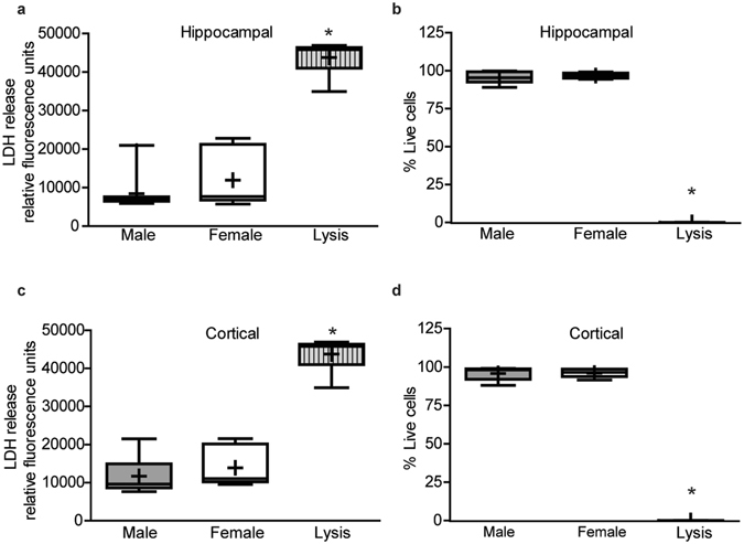
Cell viability does not differ by sex in hippocampal and cortical cultures. Sex-specific neuron-glia co-cultures were established from P0 mouse hippocampi (a,b) or neocortices (c,d). On DIV 9, cell viability was analyzed by measuring lactate dehydrogenase (LDH) release into the medium (a,c) or by quantifying the percentage of live cells in cultures stained with calcein AM (biomarker of live cells) and propidium iodide (biomarker of dead cells) using Metamorph image analysis software (b,d). As a positive control, a subset of cultures for each experimental condition were lysed with 0.2% Tritox X (Lysis). In box plots, “+” indicates the mean; whiskers, the 10–90th percentile (n = 6–9 wells per sex from at least three independent dissections). Live dead cell analysis was conducted on imaged sites (9 fields/well) which contained greater than 250 total cells counted per field. Significant differences were determined using one-way analysis of variance (ANOVA) followed by Tukey’s post hoc analysis for parametric data (b) or Kruskal Wallis test followed by Dunn’s post hoc analysis for nonparametric data (a,c,d). Asterisk indicates a significant difference between groups at p ≤ 0.05.
