Figure 7.

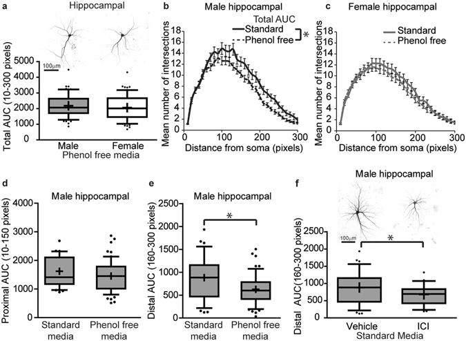
In vitro sex differences in dendritic morphology of hippocampal neurons is mediated by estrogen-dependent mechanisms. Sex-specific neuron-glia co-cultures were established from P0 mouse hippocampi, grown in standard culture medium containing phenol red or in phenol red-free medium, transfected with MAP2B-GFP plasmid on day in vitro (DIV) 6, treated with vehicle (0.05% DMSO) or estrogen receptor antagonist (ICI) on DIV 7 and fixed on DIV 9. As shown in representative images and box plots illustrating the total area under the curve (AUC) of Sholl plots of DIV 9 GFP-positive male and female hippocampal neurons (a), sex differences in dendritic morphology are not observed when neurons are cultured in medium without phenol red. Sholl plots of male (b) and female (c) hippocampal cultures grown in culture medium with or without phenol red; dendritic morphology was assessed by quantifying the total area under the Sholl curve. Dendritic morphology of male hippocampal neurons grown in culture media with or without phenol red was quantified by measuring: (d) the proximal (10–150 pixels from the soma) and (e) distal (160–300 pixels from soma) area under the curve (AUC). Representative images and Sholl analyses of distal AUC of DIV 9 GFP-positive male hippocampal neurons grown in standard culture media and treated with either vehicle or the estrogen receptor antagonist ICI (1 μM) (f). In box plots (a, d-f), “+” indicates the mean; whiskers, the 10–90th percentile (n = 23–60 neurons per sex per group from at least three independent dissections). Significant differences were determined using Student’s T-test for parametric data (a,b,c,e,f) and Mann-Whitney U test for nonparametric data (d). Asterisk indicates a significant difference between groups at p ≤ 0.05. AUC = area under the curve. Magnification; 0.65 microns per pixel.
