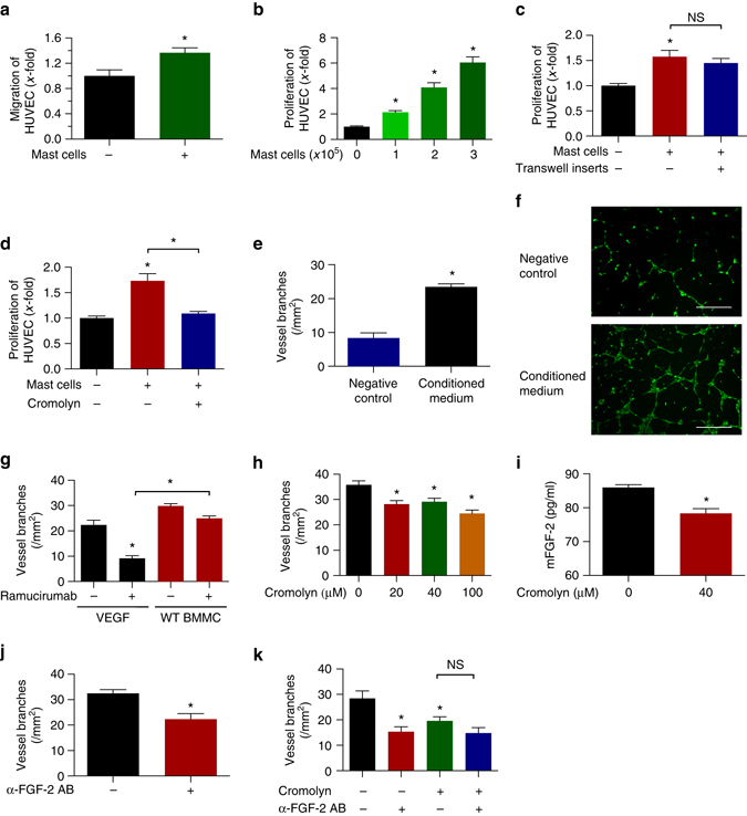
Fig. 5.

Mast cells secrete pro-angiogenic factors that alter the proliferative and organizational state of endothelial cells. a Transwell migration assay of HUVEC stimulated with MC-conditioned medium (n = 3; *p < 0.05; two-tailed t-test). b Direct co-culture of HUVEC and BMMC (n = 11–12; *p < 0.05; one-way ANOVA). c Indirect co-culture of HUVEC and BMMC (n = 3; *p < 0.05; one-way ANOVA). d HUVEC were cultured in the presence of MC–conditioned medium (n = 3; *p < 0.05; one-way ANOVA). e Tube formation assays of HUVEC in the presence of serum free or MC-conditioned medium (n = 3–4; *p < 0.05; two-tailed t-test). f Representative pictures of tube formation assays with scale bars representing 400 µm. g Tube formation of HUVEC in response to ramucirumab. VEGFA or conditioned medium from WT BMMC was used as stimulus (n = 7–10; *p < 0.05; one-way ANOVA). h BMMC-induced tube formation of HUVEC response to different concentrations of cromolyn (n = 3; *p < 0.05; one-way ANOVA). i Secretion of MC-derived FGF-2 in response to 40 µM cromolyn (n = 2; *p < 0.05; two-tailed t-test). j BMMC-induced tube formation of HUVEC in response to 0.5 µg/ml anti-FGF-2 antibody (n = 8; *p < 0.05; two-tailed t-test). k BMMC-induced tube formation of HUVEC in response to single or combinatorial treatment with 20 µM cromolyn and 0.5 µg/ml anti-FGF-2 antibody (n = 3; *p < 0.05; one-way ANOVA). Results are shown as representative means ± s.e.m
