Fig. 1.
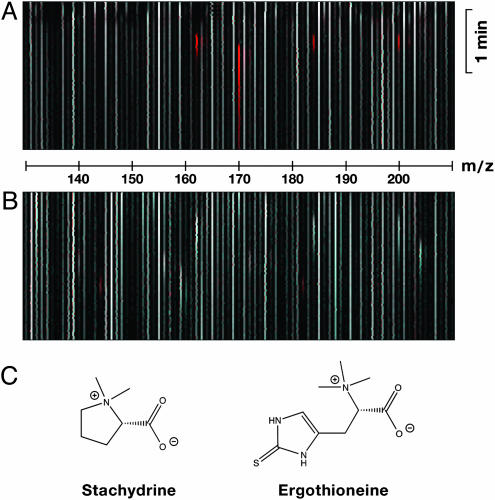
Results from LC-MS difference shading. (A) Difference image of uptake buffer plus 1-methyl-4-phenylpyridinium (m/z = 170) and carnitine (m/z = 162), each at 1 μmol/liter, with uptake buffer. Both peaks are clearly marked by red color. (B) Difference image of cell lysates. 293-FIT-ETTh cells were cultivated for 1 day in the presence (to express ETTh) or absence (control) of 1 μg/ml doxycycline in growth medium. 293 cells do not originally express OCTN1h (Fig. 8). In the uptake experiment, cells were preincubated in uptake buffer for 20 min and then incubated for 1 min with a 1:1 mixture of human plasma and uptake buffer. Cells were washed with ice-cold uptake buffer and lysed with 4 mmol/liter HClO4. For full-scan LC-MS, a m/z-range of 50–500 was recorded (scan time, 2 s). (C) Structures of stachydrine and ET.

