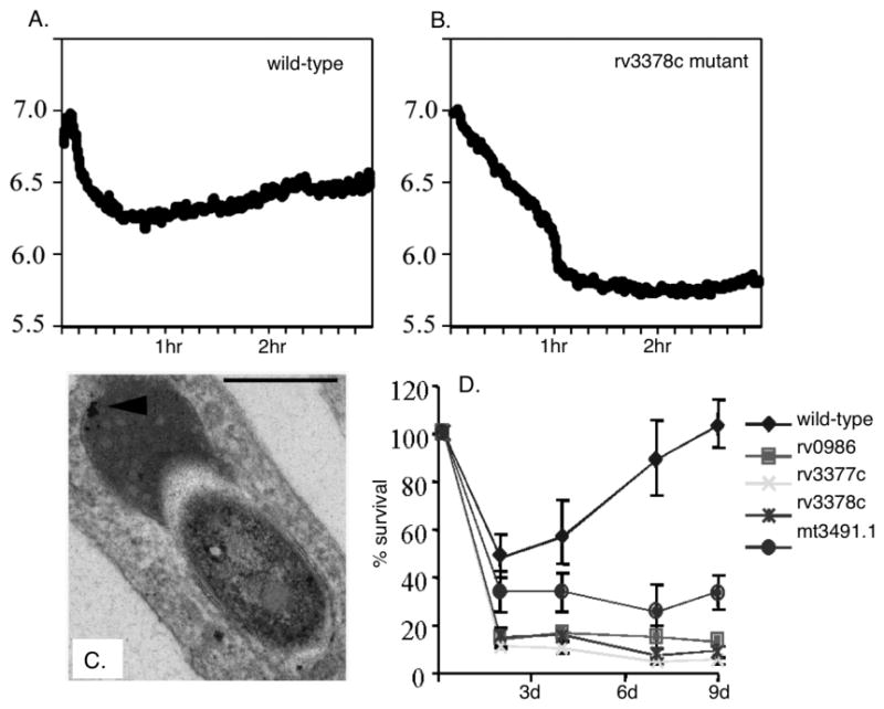
Fig. 2. Measuring the pH of Mtb-containing phagosomes.

A. Graphs showing the acidification of the phagosomes of BMMØ infected with CF-SE-labeled wild-type Mtb (5). The phagosomes containing wild-type viable bacteria acidify to pH 6.4 within the time frame of the assay (3 hrs). B. In contrast, the phagosomes containing the rv3378c mutant are able to acidify further, down to pH 5.8. C. Electron microscopy examination of macrophages infected with the mutant bacteria demonstrated that these phagosomes exhibited an increased association with lysosomes, defined by their dense cargo, and the presence of an iron dextran tracer (arrowhead). D. The survival kinetics of wild-type Mtb compared to the mutants isolated as defective in the regulation of their phagosome indicates that these mutants are unable to enter into growth phase following uptake by the BMMØ.
