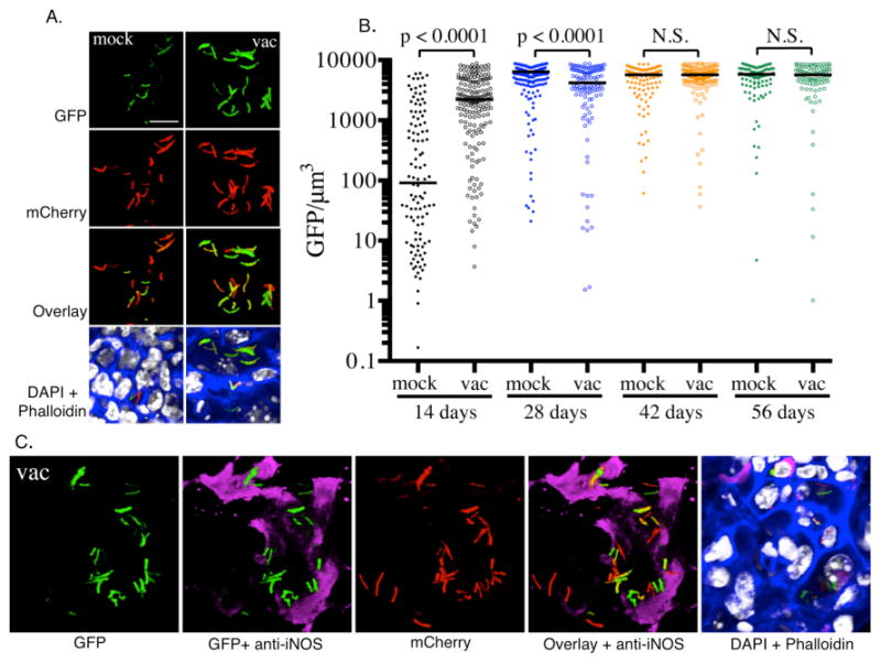
Fig. 3. Measuring the induction of hspX’-driven expression of GFP.

A. Illustrates the levels of expression of hspX’ promoter dependent GFP at 14 days post-infection in naïve mice (mock) and mice vaccinated with heat-killed Mtb (vac), as reported previously (16). At 14 days the vaccinated mice have a robust immune response, while this response is not fully developed in the naïve mice. B. Images are acquired using the Leica LAS software and are quantified by Volocity. The bacterial volume is defined by the mCherry signal, and the total GFP signal intensity is measured for each bacterial volume. Each dot on the scatter plot represents a single bacterium or a cluster of bacteria that cannot be separated. The average level of expression of GFP at 14 days is markedly lower in naïve mice than in vaccinated mice. By 28 days, high levels of induction of GFP expression is observed in both naïve and vaccinated groups. The horizontal bars represent the median value for each group and p-values were generated using a Mann-Whitney statistical test. C. The hspX promoter is regulated by the dosR regulon, which is activated by hypoxia and nitric oxide. Nitric oxide is generated by the inducible nitric oxide synthase (iNOS), which is expressed in mouse macrophages in the presence of IFN-γ and TNF-α. Probing the murine lung tissue with an anti-iNOS antibody reveals the presence of the host enzyme in regions that contain GFP-expressing Mtb.
