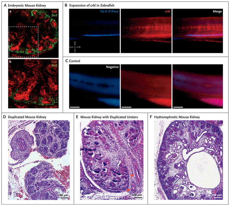
Figure 3. Localization of Crkl in Developing Urinary Tracts in Mice and Zebrafish and Phenotypes of Crkl Knockout Mice.
Panel A shows immunostaining for Crkl in kidney obtained from a transgenic mouse on embryonic day E15.5, in which Six2 has been tagged with enhanced green fluorescent protein (GFP), with specific Crkl staining of the ureteric bud (in red) surrounded by Six2-positive cap mesenchyme cells (in green) (subpanel a). A magnified field shows ureteric-bud branching within condensing metanephric mesenchyme (subpanel b). Panel B shows specific pronephros expression of crkl in zebrafish, as shown by colocalization after staining with antibody against sodium–potassium ATPase. In the orientation symbol, D denotes dorsal, V ventral, C caudal, and R rostral. Panel C shows images of negative controls (i.e., fish treated with fluorophore-conjugated secondary antibodies only). In Panels B and C, the scale bars represent 100 μm. In a mouse model that targets Crkl exon 2, three crosses with transgenic Cre-recombinase mice were created to effect the deletion of exon 2 in specific compartments: E2a-Cre for global knockout, Six2-Cre in the cap mesenchyme, and Hoxb7 in the structures derived from ureteric buds. Panel D shows tissue from a Six2-Cre mouse in which duplication of the right kidney is accompanied by an irregular, dysplastic pattern or ureteric-bud branching on embryonic day E15.5. Panel E shows tissue from an E2a-Cre mouse in which a single kidney with duplicated ureters (arrowheads) is accompanied by failure of medullary and renal papillary development on day E14.5. Panel F shows tissue from a Six2-Cre mouse, in which the kidney is hydronephrotic with dilated pelvis, absence of medullary architecture, and several microcystic glomeruli and tubules on day E15.5.

