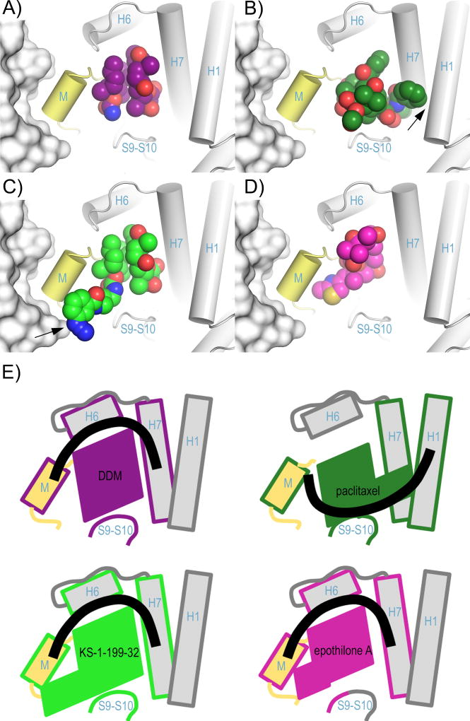
Figure 4. Binding of taxane-site ligands in the context of the microtubule.
Superposition of the intermediate domains of (A) DDM, (C) KS-1-199-32 and (D) EpoA (PDB ID 4I50) onto the corresponding domain of (B) the paclitaxel-bound microtubule (PDB ID 3J6G). The helices shaping the taxane site of the microtubule are displayed as cylinders, the β-tubulin subunits of the flanking protofilament are in surface representation. The M-loop is highlighted in yellow. The ligands are in spheres representation. Key interactions are marked with a black arrow. (E) Schematic representation of the structural features shown in panels (A) – (D) highlighting the secondary structural elements contacted by the individual ligands. The ligand contacted elements are framed with the same color code as the individual ligands. The thick black lines denote the connection between the secondary structural elements.

