Figure 4.
Subcellular Localization of AtCPT7.
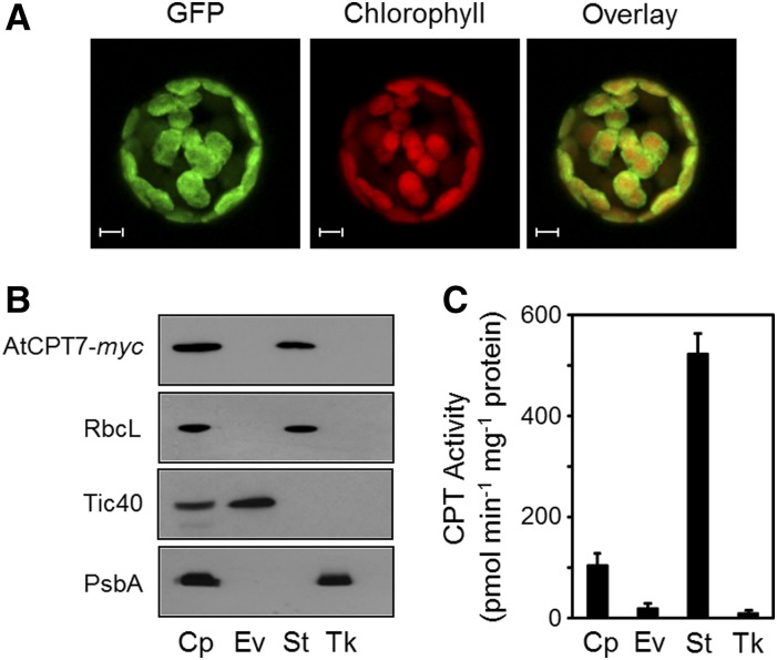
(A) Protoplasts were prepared from Arabidopsis plants stably expressing GFP fused to the C terminus of AtCPT7. Fluorescence attributable to GFP, chlorophyll, and their merged signals were observed by confocal microscopy. Bars = 5 µm.
(B) Fractionation of chloroplasts from Arabidopsis plants stably expressing AtCPT7-myc. Intact chloroplasts (Cp) were fractioned into envelope (Ev), stroma (St), and thylakoid (Tk) compartments by sucrose gradient centrifugation. Protein samples from each fraction were resolved by SDS-PAGE and analyzed by immunoblotting using antibodies specific for the myc tag of AtCPT7, the Rubisco large subunit (RbcL), the Tic40 component of the inner envelope translocon, or the D1 reaction center protein (PsbA) of PSII.
(C) CPT enzyme activity in chloroplasts. Intact chloroplasts from wild-type Arabidopsis plants were fractioned as in (B), and each compartment was assayed for CPT enzyme activity using GGPP and 14C-IPP as substrates. Enzymatic products were analyzed and quantified as described in Methods, and the data represent the means ± sd from three independent experiments.

