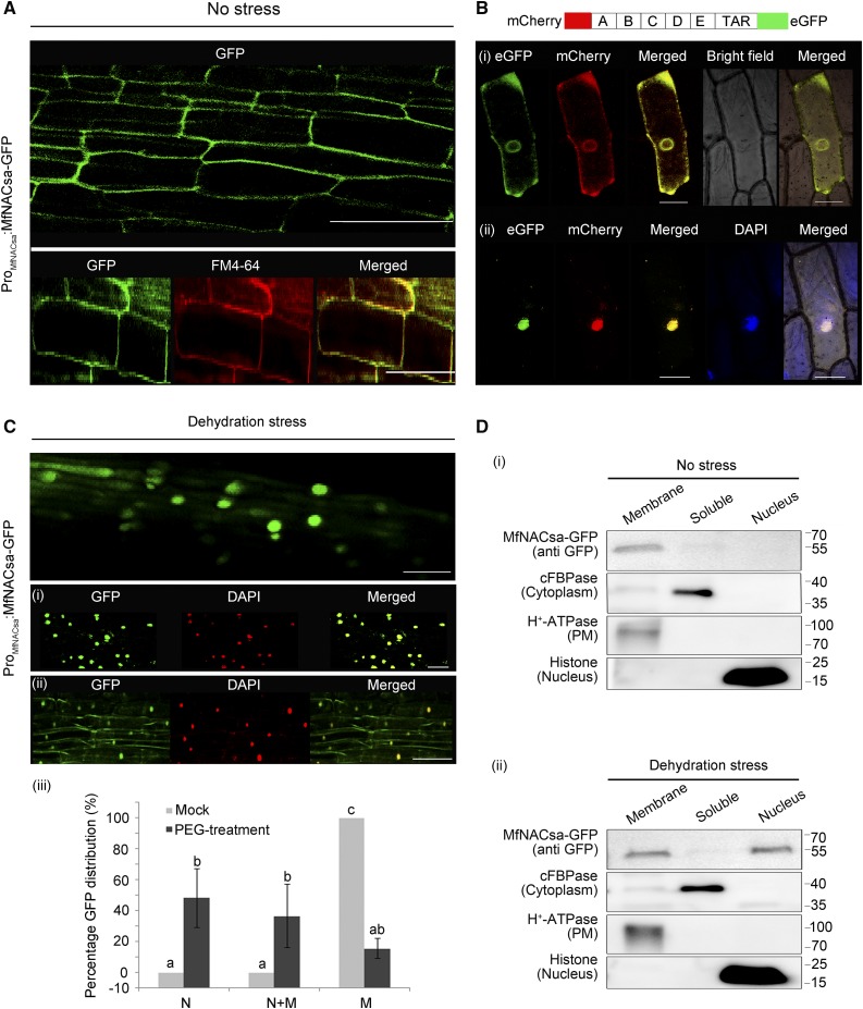
Figure 2.
MfNACsa Is Translocated to the Nucleus under PEG-Imposed Dehydration Stress.
(A) Transgenic M. truncatula hairy roots expressing the MfNACsa-GFP fusion proteins driven by native promoter were stained with the membrane dye FM4-64 for 5 min under normal conditions (no stress), with an ICQ of 0.114.
(B) After hairy roots transiently expressing MfNACsa-GFP driven by the native promoter were treated with 50% PEG-8000 for 2 h (dehydration stress), the steady state locations of MfNACsa-GFP protein were imaged and stained with DAPI dye (pseudocolor, red; [i] and [ii]). Bars = 50 μm. The percentage of cells expressing GFP at the nucleus (N), nucleus and membranes (N+M), or membranes (M) in transgenic hairy root cells were counted respectively (iii). The data represent the mean and se of three independent replicates (n > 15) (nonparametric one-way ANOVA and Duncan multiple range test, P < 0.05).
(C) Nucleus translocation of the mCherry-MfNACsa-eGFP fusion protein under 10% PEG-8000 treatment in onion epidermal cells. The mCherry and eGFP signal patterns were observed at 0 (i) and 2 h (ii) after 10% PEG-8000 treatment by confocal imaging (Olympus FluoView FV1000). The same cells were imaged before and after treatment. Bars = 50 μm.
(D) Cellular fractionation and immunoblot detection of transiently expressed ProMfNACsa:MfNACsa-GFP in N. benthamiana leaves under normal conditions (i) or 30% PEG-8000 treatment for 2 h (ii). MfNACsa-GFP is shown. cFBPase, H+-ATPase, and Histone H3 mark the cytoplasm, PM, and nuclear fraction, respectively.

