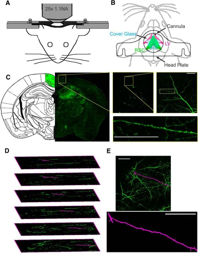
Figure 1.

In vivo multiphoton imaging of dendritic spines. A, Schematic representation of custom stage mount to stabilize mouse for multiphoton imaging. B, Diagram of location of head plate (outline), ICV-directed guide cannula (black), and glass coverslip (blue) in relation to RSC (green) and lateral ventricle (LV, magenta). C, Location and example dendrite from fixed Thy1-GFP brain section showing source of spine images in RSC agranular cortex. D, Expanded image stack demonstrating dendrite extraction (magenta) from a complex 3D structure (green). E, Extracted dendrite segment (magenta) shown as an overlay on max-Z projection of image stack (green) and as individual image. Scale bars, 50 μm.
