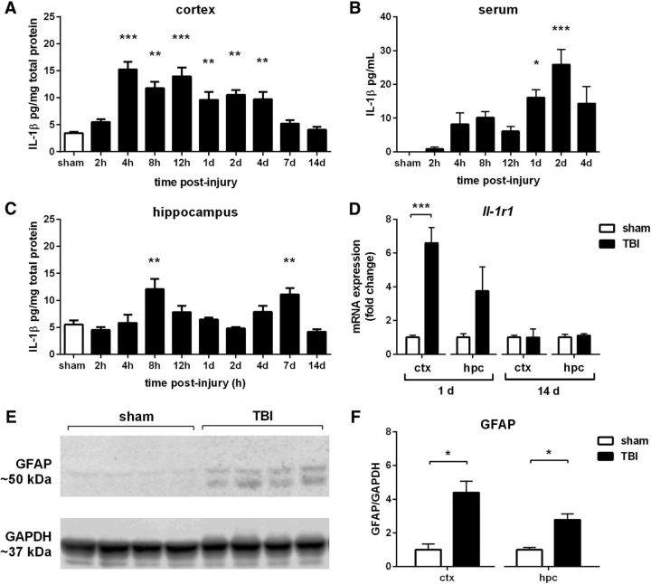
Figure 2.
IL-1 response after pTBI. IL-1β protein was detected by ELISA across a time course after pTBI, in the ipsilateral cortex (A), hippocampus (C), and serum (B; one-way ANOVA with Dunnett's post hoc). Quantitative PCR revealed a significant increase in Il-1r1 receptor in the cortex (D) at 1 d post-injury, with a trend in the hippocampus (p = 0.09). GFAP, an indicator of astrocyte activation detected by Western blot (E), was also elevated at 1 d post-injury compared with sham controls, in both the injured cortex and hippocampus (F). *p < 0.05, **p < 0.01, ***p < 0.001; n = 4–6/group.

