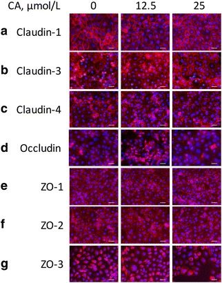
Fig. 4.

The distributions of TJ proteins claudin-1 (a), claudin-3 (b), claudin-4 (c), occludin (d), ZO-1 (e), ZO-2 (f), and ZO-3 (g) in IPEC-1 cells. Cells were treated as in Fig. 3, and immunofluorescence staining was performed to identify the distributions of the proteins. CA, Cinnamicaldehyde; IPEC-1, intestinal epithelial porcine cell line 1. Scale bar, 50 μm
