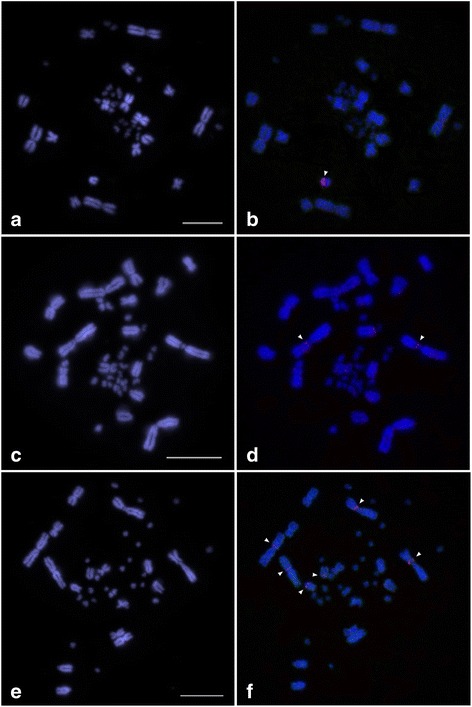
Fig. 3.

Chromosomal distribution of the VSAREP1 and VSAREP2 sequences on a DAPI-stained metaphase spread prepared from three Australian varanid lizards: Varanus acanthurus (a, b), V. gouldii (c, d), and V. rosenbergi (e, f). Hybridization patterns of Spectrum Orange-labeled VSAREP1 (red) (b, d, f) and SpectrumGreen-labeled VSAREP2 (green) (no signal) on DAPI-stained chromosomes. Fluorescent DAPI-stained pattern of chromosomes are shown in a, c, and e. Arrowheads indicate the hybridization signals. Scale bar represents 10 μm. VSAREP1 sequences were localized to the largest microchromosome in V. acanthurus, at the pericentromeric region of chromosome 1p in V. gouldii, and at the pericentromeric regions of chromosome 1p and 2p and the centromeric region of chromosome 7 in V. rosenbergi
