Fig. 4.

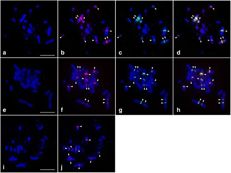
Chromosomal distribution of the VSAREP satellite DNA (stDNA) isolated from each Australian varanids on a DAPI-stained metaphase spread prepared from three Australian varanid lizards: Varanus acanthurus (a – d), V. rosenbergi (e – h), and V. gouldii (i – j). Hybridization patterns of rhodamine-labeled VSAREP stDNA (red) ((V. acanthurus, clone no. 3: SFII) b, (V. rosenbergi, clone no. 14: SFII) f, and (V. gouldii, clone no. 13: SFI) j) or FITC-labeled VSAREP stDNA (green) ((V. acanthurus, clone no. 4: SFIII) c and (V. rosenbergi, clone no. 9: SFI) g) and their co-hybridization pattern (d, h). Fluorescent DAPI-stained pattern of chromosomes are shown in a, e, and i. Arrowheads indicate the hybridization signals. Scale bars represent 10 μm
