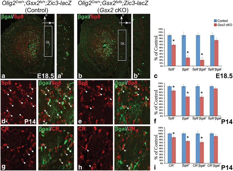
Fig. 5.

Impairment of septum-derived PGCs in the Gsx2 cKO OB. (a-c) Septum-derived GL cells marked by βgal were reduced, leading to a significant loss of septum-originated Sp8+ interneurons (i.e. βgal+) and a milder reduction of total Sp8+ PGCs in the E18.5 Gsx2 cKO OB, whereas LGE-derived Sp8+ interneurons (i.e. βgal−) were largely normal. Boxes in (a) and (b) indicate the GL area shown in (a’) and (b’) respectively. (d-f) Sp8+ PGCs were reduced in the P14 Gsx2 cKO OB, primarily due to the reduced number of the septum-derived (i.e. βgal+) interneurons. Arrowheads indicate Sp8+βgal+ cells that originated from the septum. (g-i) P14 Gsx2 cKO OB showed reduced number of CR+ PGCs, particularly those βgal-expressing ones generated from the septum. Other CR+ PGCs (i.e. βgal−), presumably originating from other regions including LGE, were largely intact. Data represent the mean ± s.e.m. *p < 0.05
