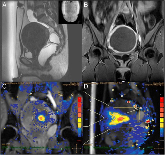
Fig. 2.

A 33-year-old woman with 10.1-cm uterine fibroid, without an abdominal scar. a Sagittal T2W planning MR image of uterine fibroid prior to MRgHIFU treatment. b CE-T1W image acquired immediately after MR-guided high-intensity focused ultrasound treatment. The NPV ratio was 100%. c, d An example of multiplane MR thermometry acquired in both coronal and sagittal planes during sonication
