
Fig. 5.
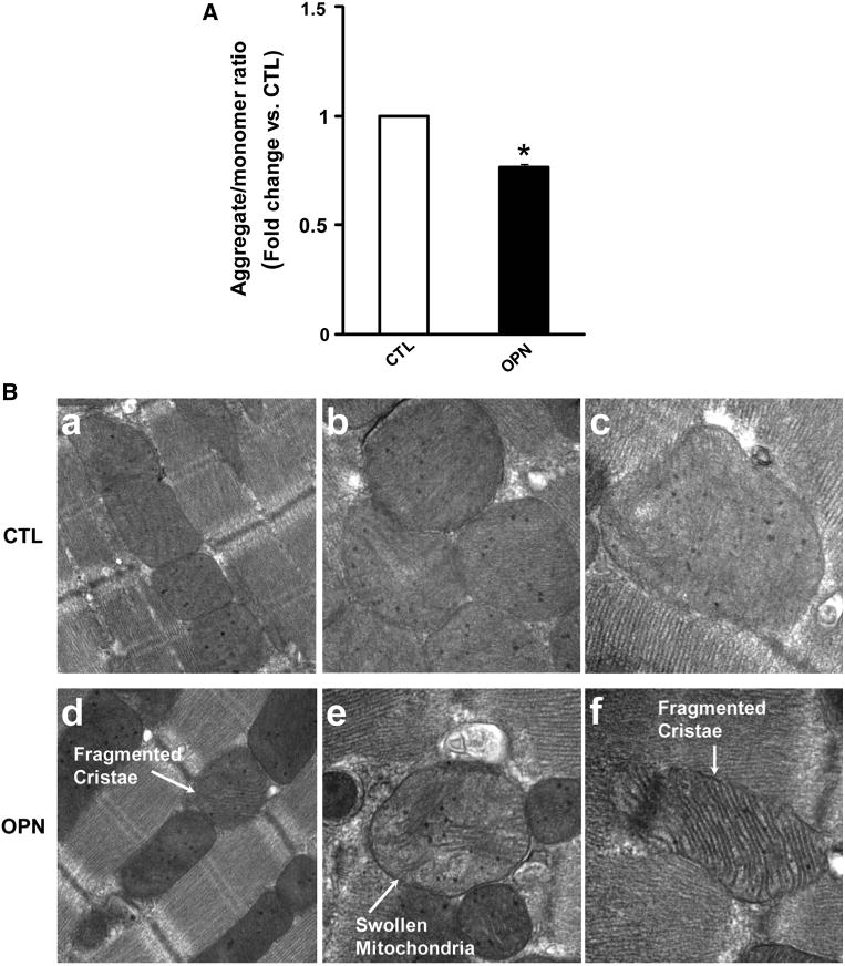
OPN induces mitochondrial depolarization and remodeling. a ARVMs were treated with OPN for 6 h and incubated with JC−1 dye for 10 min at 37 °C. The cells were visualized using epifluorescence microscope and the ratio of red-to-green fluorescence was calculated. *p < 0.05 versus CTL; n = 3. b ARVMs were treated with OPN for 48 h. After fixing and processing, the cells were visualized using transmission electron microscope. Photomicrographs illustrate ultrastructural features of mitochondria in control (CTL; a ×944,000; b, c ×973,000) and OPN-treated (d ×44,000; e, f × 73000) ARVMs