
Fig. 6.
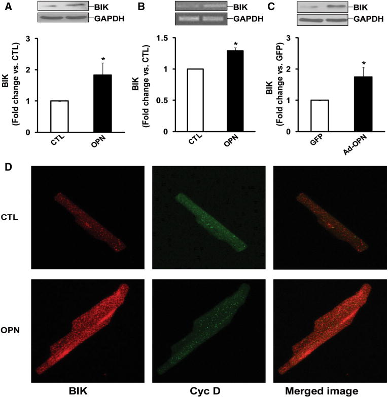
OPN increases BIK expression. a ARVMs were treated with OPN for 24 h. Total cell lysates were analyzed by Western blot using anti-BIK antibodies. The lower panel exhibits the mean data normalized to GAPDH, *p < 0.05 versus CTL; n = 9. b ARVMs were treated with OPN for 24 h. Total RNA were reverse transcribed and resulting cDNAs were subjected for amplification of BIK and GAPDH genes, *p < 0.05 versus CTL; n = 4. c ARVMs were infected with adenoviruses expressing OPN or GFP for 48 h. Cell lysates were analyzed by Western blot using anti-BIK antibodies. The lower panel exhibits the mean data normalized to GAPDH, *p < 0.05 versus CTL; n = 5. d ARVMs were treated with OPN for 24 h and immunostained using anti-BIK and anti-cyclophilin D antibodies. Cells were visualized using confocal microscope. The figure depicts representative images from three independent experiments