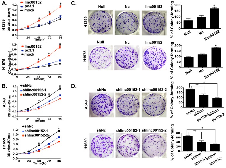
Figure 3.
Linc00152 stimulated tumor cell proliferation in lung adenocarcinoma. A: The CCK8 cell counting assays revealed the living cell growth curves of H1299 and H1975 cells that were transfected with the indicated plasmids at the indicated time points. Error bars are shown. *: p<0.01. B: The CCK8 cell counting assays revealed the living cell growth curves of A549 and H1650 cells that were infected with the indicated lenti-viruses at the indicated time points. Error bars are shown. *: p<0.01. C: Representative images of the colony-forming results of H1299 and H1975 cells that were infected with the indicated lenti-virus for 14 days. *: p<0.01. D: Representative images of the colony-forming results of A549 and H1650 cells that were infected with the indicated lenti-virus for 14 days. *: p<0.01. **: p<0.05.

