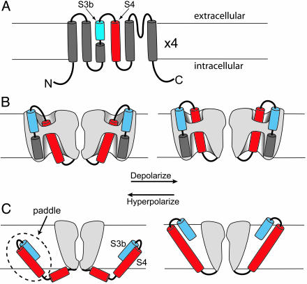
Fig. 1.
Models of topology and voltage sensor movement. The membrane is indicated by horizontal lines. (A) Topology of a potassium channel subunit with six transmembrane segments (S1–S6). The S3 segment is divided into two helices, S3a and S3b. The S4 segment is in red. (B) Conventional model of voltage sensor movement. Two of the four subunits are shown (sliced open to expose the “gating pore”) with the ion permeation path between them. The foreground and background subunits are removed for clarity. Gray represents protein. Depolarization (Right) moves the extracellular portion of the S4 segment (red) outward through a short hydrophobic gating pore, opening the permeation pathway. Most of the S4 segment is surrounded by hydrophilic vestibules. The transmembrane electric field falls mainly across the gating pore. (C) Paddle model, which is adapted from ref. 4. Two opposing subunits are shown. Depolarization moves the paddle outward through lipid, pulling the cytoplasmic activation gate open. The transmembrane electric field felt by S4 arginines falls mainly across the lipid. B and C are adapted from ref. 11.

