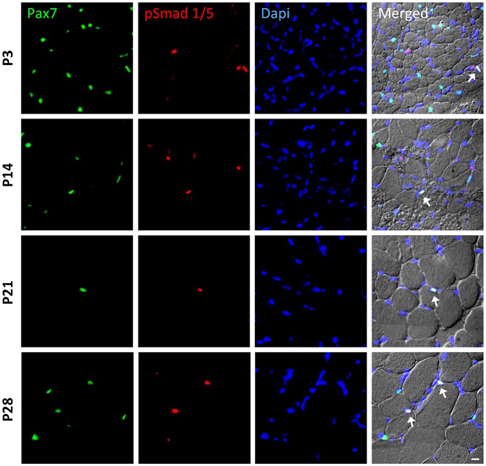
Fig. 1.
BMP signaling activity in satellite cells during postnatal muscle growth. Sample images of immunohistochemistry to monitor phosphorylated Smad1/5 (p-Smad1/5) expression (red) in Pax7-expressing satellite cells (green). Muscle sections were obtained from the tibialis anterior (TA) muscle of postnatal wild-type mice at P3, P14, P21 and P28 (panels from top to bottom). DAPI (blue) was used as a nuclear stain. All three channels (green, red and blue) were merged together with a differential interference contrast image (DIC), in which the individual fibers can be visualized. Cells that co-express p-Smad1/5 and Pax7 are highlighted with arrows. Scale bar: 50 µm.

