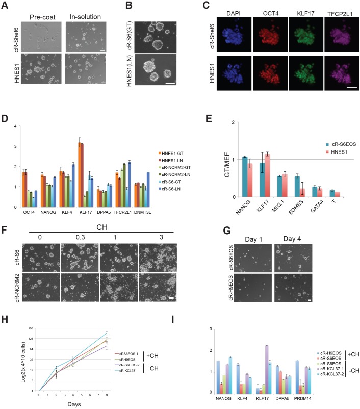
Fig. 2.
Feeder-free culture. (A) Cells plated on Geltrex-coated plates (left) or with Geltrex added to the medium (right). Images taken after 4 days. (B) Cultures in Geltrex (GT) or laminin (LN) for more than ten passages. (C) Immunostaining for pluripotency markers in reset cells passaged in laminin. (D) Naïve marker expression in feeder-free reset cultures in t2iLGö as determined by RT-qPCR and normalised to the expression level in H9-NK2 transgene reset cells. (E) Lineage marker expression in feeder-free reset cultures relative to levels on feeders. (F) Reset cells plated in the presence of the indicated concentrations (µM) of the GSK3 inhibitor CHIR99021 (CH) for 4 days. (G) Images of colony expansion over 4 days in Geltrex. (H) Growth curve for reset cells in tt2ilGö and Geltrex. Error bars indicate s.d. from triplicate cultures. (I) RT-qPCR marker profile for cells reset with or without CH and expanded in tt2iLGö and Geltrex, normalized to expression level in cR-H9 cells on MEF in tt2iLGö. Error bars on PCR plots indicate s.d. of technical duplicates. Scale bars: 100 μm in A,B,F,G; 50 μm in C.

