
Figure 2. Crystal Structure of Human Pds5B Bound to YSR motif of Wapl.
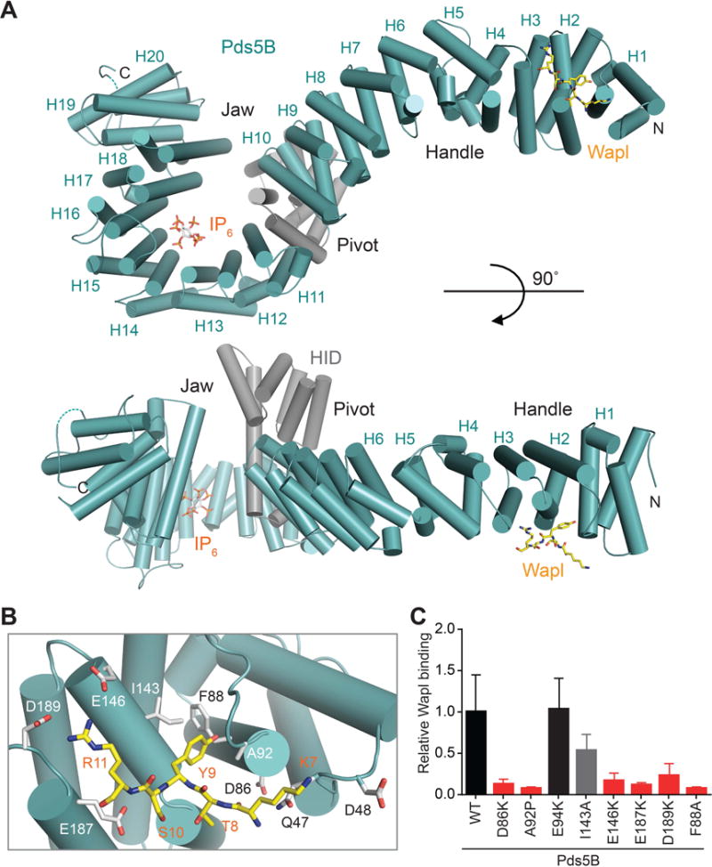
(A) Cartoon drawing of the crystal structure of human Pds5B in complex with the YSR motif of Wapl in two different orientations. The HEAT repeats and the helical insert domain (HID) are colored teal and gray, respectively. The Wapl peptide and IP6 are shown as sticks. The N- and C-termini and the 20 HEAT repeats (H1–H20) are labeled. Pds5B is shaped like a plier lever, with H1–H8 resembling the handle, the HID resembling the pivot, and H9–H20 forming the jaw. All structure figures are made with PyMOL (www.pymol.org).
(B) Zoomed in view of the Pds5B–Wapl interface. Pds5B and Wapl residues are shown as gray and yellow sticks, respectively.
(C) Quantification of the relative 35S-Pds5B intensities bound to GST-Wapl1–150. Error bars, SD (n = 3 independent experiments) (see also Figures S2 and S3).