
Figure 4.
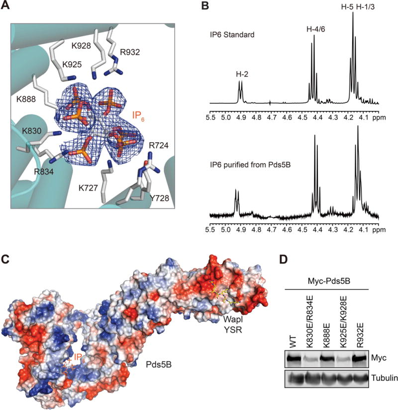
IP6 as a Structural Cofactor of Pds5B
(A) Zoomed in view of the IP6-binding site of Pds5B. IP6 is shown as stick, along with its 2Fo-Fc electron density map (blue mesh) contoured at 1.0 σ. IP6-binding residues are shown as sticks and labeled.
(B) 1D 1H NMR spectra of authentic IP6 standard (top) and IP6 isolated from recombinant human Pds5B, with the 1H assignment indicated.
(C) Surface drawing of human Pds5B colored with its electrostatic potential (blue, positive; red, negative). IP6 and the Wapl peptide are shown in sticks.
(D) Anti-Myc and anti-β-tubulin immunoblots of lysates of HeLa cells transfected with the same amount of the indicated Myc-Pds5B plasmids. WT, wild type (see also Figure S6).