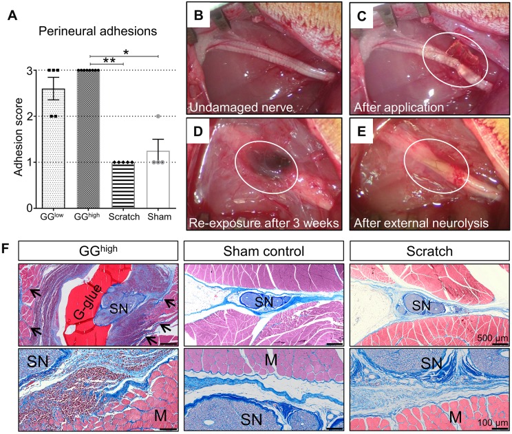
Fig. 1.
Analysis of perineural adhesions between the sciatic nerve and the surrounding tissue 3 weeks after primary surgery. (A) Rats belonging to the GGhigh group (n=8) exhibited distinct adhesive fibrotic tissue requiring sharp dissection during external neurolysis, whereas sham (n=4, P=0.0125) and scratch (n=5, P=0.0012) groups had no or mild adherence . Rats of the GGlow group (n=5) also showed predominantly severe adhesions. The difference between the GGlow group and the other groups was, however, not statistically significant (P=0.2129 and P=0.0562). *P<0.05, **P<0.01. (B-E) Morphological gross evaluation of the sciatic nerve. Circled area shows the sciatic nerve immediately after glue application (C), 3 weeks afterwards during re-exposure (D) and after removal of the glue and the developed adhesions (E; external neurolysis). (F) Histological en bloc cross sections of GGhigh, sham control and scratch groups stained with CAB, showing the sciatic nerves and surrounding tissue 3 weeks following primary surgery. The application of glutaraldehyde glue induced strong inflammatory mononuclear cell infiltration of both nerve and muscles, as well as severe growth of dense collagenized matrix infiltrating muscle fibres (arrows). Although, in the scratch group, a slight increase of loose connective tissue could be observed, the surrounding muscles were not affected, similar to sham controls, which showed only mild formation of collagen. G-glue, glutaraldehyde glue; SN, sciatic nerve; M, muscle fibres.

